By Patrick R. Gavin
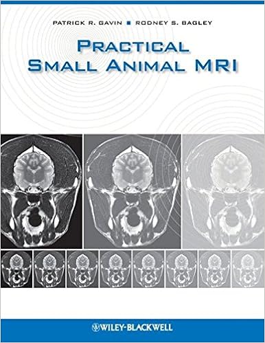
Useful Small Animal MRI is the seminal reference for clinicians utilizing Magnetic Resonance Imaging within the analysis and therapy of veterinary sufferers. even though MRI is used most often within the analysis of neurologic problems, it additionally has major software to different physique structures. This ebook covers general anatomy and particular scientific stipulations of the frightened procedure, musculoskeletal approach, stomach, thorax, and head and neck. It additionally comprises numerous chapters on affliction of the mind and backbone, together with inflammatory, infectious, neoplastic, and vascular illnesses, along congenital and degenerative issues.
Read or Download Practical Small Animal MRI PDF
Similar diagnostic imaging books
Image-Processing Techniques for Tumor Detection
Univ. of Arizona, Tucson. presents a present assessment of laptop processing algorithms for the id of lesions, irregular lots, melanoma, and disorder in clinical photographs. provides examples from various imaging modalities for larger reputation of anomalies in MRI, CT, SPECT, and digital/film X ray.
Coronary Artery CTA: A Case-Based Atlas
Coronary Artery CTA: A Case-Based Atlas offers the reader with a evaluation of a extensive variety of cardiac CT angiography (CCTA) situations from the instructing dossier of Dr. Claudio Smuclovisky. every one case contains vast CCTA photos, a short historical past, analysis, dialogue, and pearls and pitfalls. The target of the e-book is to supply the reader with a huge diversity of CCTA situations that come with basic anatomy, congenital coronary anomalies, coronary artery affliction, percutaneous coronary intervention, postsurgical coronary revascularization, and extra-coronary abnormalities.

Electron transfer reactions: inorganic, organometallic, and biological applications
Starts off with a old assessment by means of Henry Taube. Overviews the advances pioneered by means of Taube, together with mechanisms of electron move reactions, cost move complexes, and *p again bonding results in metal-ligand interactions. Discusses purposes of ideas of electron move to diversified parts of chemistry and biology corresponding to the selective and regulated oxidation of natural practical teams, polymerization catalysis, steel organic interactions with DNA, organic electron move reactions, and new imaging brokers in diagnostic drugs.
Introduction to the Science of Medical Imaging
Innovative advances in imaging know-how that supply excessive answer, 3-D, non-invasive imaging of organic matters have made biomedical imaging a vital device in scientific medication and biomedical study. Key technological advances contain MRI, positron emission tomography (PET) and multidetector X-ray CT scanners.
- PET : physics, instrumentation, and scanners
- Case Studies in Abdominal and Pelvic Imaging
- Computed Tomography of the Coronary Arteries
- Coronary Radiology (Medical Radiology Diagnostic Imaging)
- Neuroimaging Personality, Social Cognition, and Character
- Clinical Cardiac MRI
Extra resources for Practical Small Animal MRI
Example text
The strength of the gradient coils determines the ability of an MR unit to change the magnetic field per unit distance. The strength of the gradient coils will have the largest contribution to end-plane resolution. The RF coil, besides an emitting RF coil, is also a receiving coil capable of detecting the signal from the spins within the body. Most modern magnets are self-shielded with an opposing magnetic field such that they do not need to be magnetically shielded. All magnets do, however, need to have some form of RF shielding.
Transverse T2-weighted (A) and T1-weighted (B) MR images from a dog at the level of the frontal sinus. Note the hypointense signal from air in these cavities (arrows). Sagittal T2-weighted (C) and T1-weighted (D), and dorsal T1-weighted (E) MR images from a dog with an air-filled frontal sinus. 23. Sagittal T1-weighted MR images from two separate dogs, one with (A) and without (B) and air-filled frontal sinus. Sagittal T2-weighted MR image (C) from a brachycephalic dog breed without an air-filled frontal sinus (arrow).
Transverse proton density MR image (C) and gross brain image (D) from a dog at the level of the caudal thalamus and geniculate bodies. Transverse T2-weighted MR image (E) and gross brain image (F) from a dog at the level of the caudal geniculate bodies and rostral mesencephalon. 11. Transverse (A) and sagittal (B), T1-weighted images prior to intravenous contrast administration from a dog showing the focal, hyperintense signal from the pituitary (arrows). thalamus. From here, visual fibers travel further caudally in the optic radiations to synapse in the visual cortex of the occipital hemisphere.