By Miles N. Wernick, John N. Aarsvold
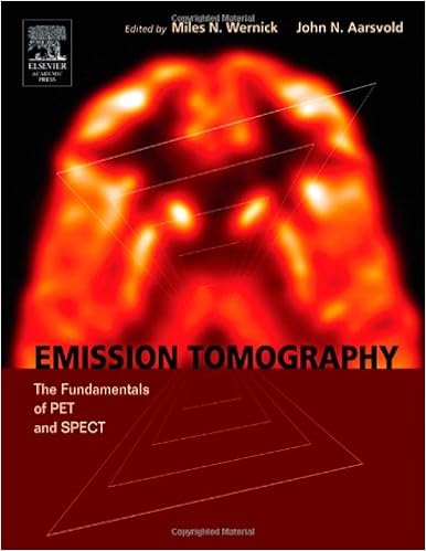
Puppy and SPECT are of contemporary most crucial medical-imaging equipment, offering pictures that show refined information regarding physiological approaches in people and animals. Emission Tomography: the basics of puppy and SPECT explains the physics and engineering rules of those very important functional-imaging tools. The expertise of emission tomography is roofed intimately, together with old origins, clinical and mathematical foundations, imaging structures and their parts, photograph reconstruction and research, simulation strategies, and scientific and laboratory purposes. The publication describes the cutting-edge of emission tomography, together with all aspects of traditional SPECT and puppy, in addition to modern subject matters resembling iterative photograph reconstruction, small-animal imaging, and PET/CT structures. This e-book is meant as a textbook and reference source for graduate scholars, researchers, scientific physicists, biomedical engineers, engineers and physicists within the medical-imaging undefined. Thorough tutorials of primary and complicated subject matters are awarded through dozens of the best researchers in puppy and SPECT. SPECT has lengthy been a mainstay of medical imaging, and puppy is now one of many world's quickest turning out to be scientific imaging recommendations, due to its dramatic contributions to melanoma imaging and different functions. Emission Tomography: the basics of puppy and SPECT is a vital source for realizing the expertise of SPECT and puppy, the main customary sorts of molecular imaging. *Contains thorough educational remedies, coupled with assurance of complex themes *Three of the 4 holders of the distinguished Institute of electric and Electronics Engineers scientific Imaging Scientist Award are bankruptcy participants *Include colour art
Read or Download Emission Tomography: The Fundamentals of PET and SPECT PDF
Similar diagnostic imaging books
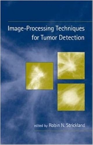
Image-Processing Techniques for Tumor Detection
Univ. of Arizona, Tucson. offers a present assessment of computing device processing algorithms for the id of lesions, irregular lots, melanoma, and ailment in clinical photographs. provides examples from a number of imaging modalities for larger attractiveness of anomalies in MRI, CT, SPECT, and digital/film X ray.
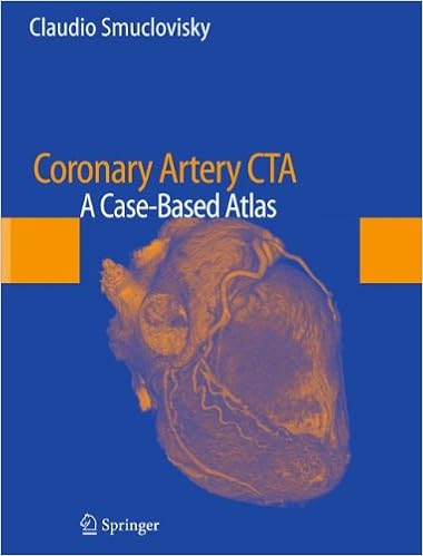
Coronary Artery CTA: A Case-Based Atlas
Coronary Artery CTA: A Case-Based Atlas provides the reader with a evaluation of a huge variety of cardiac CT angiography (CCTA) situations from the instructing dossier of Dr. Claudio Smuclovisky. every one case comprises large CCTA pictures, a short background, prognosis, dialogue, and pearls and pitfalls. The target of the ebook is to supply the reader with a wide diversity of CCTA circumstances that come with general anatomy, congenital coronary anomalies, coronary artery illness, percutaneous coronary intervention, postsurgical coronary revascularization, and extra-coronary abnormalities.

Electron transfer reactions: inorganic, organometallic, and biological applications
Starts off with a old evaluate by way of Henry Taube. Overviews the advances pioneered through Taube, together with mechanisms of electron move reactions, cost move complexes, and *p again bonding results in metal-ligand interactions. Discusses functions of ideas of electron move to different components of chemistry and biology similar to the selective and regulated oxidation of natural useful teams, polymerization catalysis, steel organic interactions with DNA, organic electron move reactions, and new imaging brokers in diagnostic medication.
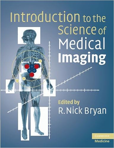
Introduction to the Science of Medical Imaging
Progressive advances in imaging expertise that offer excessive solution, 3-D, non-invasive imaging of organic topics have made biomedical imaging a vital software in medical medication and biomedical study. Key technological advances comprise MRI, positron emission tomography (PET) and multidetector X-ray CT scanners.
- Radiology of Infectious Diseases: Volume 1
- Imaging of the Cervical Spine in Children
- Neuroimaging in child neuropsychiatric disorders
- Handbuch diagnostische Radiologie: Muskuloskelettales System 3
- Diagnostic Imaging: Orthopaedics
Additional info for Emission Tomography: The Fundamentals of PET and SPECT
Example text
There are two principal alternatives to a static imaging study, dynamic studies and gated studies, both of which produce a sequence of images that can be viewed as a video. A dynamic study, which is directly analogous to an ordinary movie or video, aims to capture the radiotracer distribution as a function of time. Dynamic studies are used in visual assessments of organ function and are required for most kinds of kinetic parameter estimation. In a gated study the data acquisition is synchronized to the rhythm of the patient’s cardiac or breathing cycles.
IV. CROSS-SECTIONAL IMAGES Static ET studies aim to measure the spatial distribution of the radiotracer, which can be described by a function f (x, y, z), where, for the moment, let us define the (x, y, z) coordinates as follows with respect to a person who is standing. Let the x coordinate correspond to the left-right axis, the y coordinate to the forward-backward (anteriorposterior) axis, and the z coordinate to the up-down (superior-inferior) axis. Gated and dynamic studies add the extra dimension of time, thus producing 4D images, which can be denoted by f (x, y, z, t).
Introduction to Emission Tomography (a) (b) High counts p(xr,φ,z = 0) p(xr,φ,z = 0) y y xr xr yr yr φ φ x x Low counts FIGURE 10 (a) Idealized forward projection of image slice for a particular angle and (b) backprojection for the same angle. Bright areas of the image and projection denote high count rates, as indicated by the color bar at far left. Actual PET and SPECT data are not well described by this simplified projection model because of attenuation, blur, scatter, and noise; however, the model does capture the essential process of forming an image from projection data.