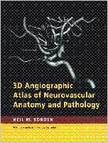
By Neil M. Borden MD
The 1st atlas to provide neurovascular info and pictures in response to catheter 3D rotational angiographic reports. The amazing 3D photos are commonly categorised and juxtaposed with traditional second angiograms for orientation and comparability. Anatomical colour drawings and concise descriptions of the most important intracranial vascular territories extra improve figuring out of the advanced cerebral vasculature. This atlas is an necessary reference for an individual looking a fuller appreciation of intracranial and cervical anatomy and pathology, despite distinctiveness.
Read Online or Download 3D Angiographic Atlas of Neurovascular Anatomy and Pathology PDF
Similar diagnostic imaging books

Image-Processing Techniques for Tumor Detection
Univ. of Arizona, Tucson. offers a present evaluation of laptop processing algorithms for the identity of lesions, irregular lots, melanoma, and affliction in scientific photos. provides examples from quite a few imaging modalities for greater attractiveness of anomalies in MRI, CT, SPECT, and digital/film X ray.
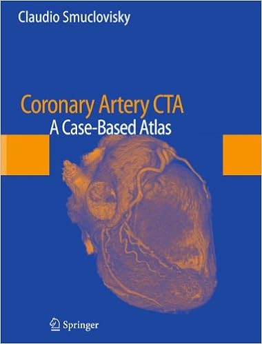
Coronary Artery CTA: A Case-Based Atlas
Coronary Artery CTA: A Case-Based Atlas offers the reader with a assessment of a large diversity of cardiac CT angiography (CCTA) instances from the educating dossier of Dr. Claudio Smuclovisky. each one case comprises broad CCTA photos, a quick heritage, analysis, dialogue, and pearls and pitfalls. The aim of the publication is to supply the reader with a large variety of CCTA circumstances that come with general anatomy, congenital coronary anomalies, coronary artery affliction, percutaneous coronary intervention, postsurgical coronary revascularization, and extra-coronary abnormalities.

Electron transfer reactions: inorganic, organometallic, and biological applications
Starts with a ancient assessment through Henry Taube. Overviews the advances pioneered through Taube, together with mechanisms of electron move reactions, cost move complexes, and *p again bonding results in metal-ligand interactions. Discusses purposes of ideas of electron move to various parts of chemistry and biology equivalent to the selective and regulated oxidation of natural sensible teams, polymerization catalysis, steel organic interactions with DNA, organic electron move reactions, and new imaging brokers in diagnostic medication.
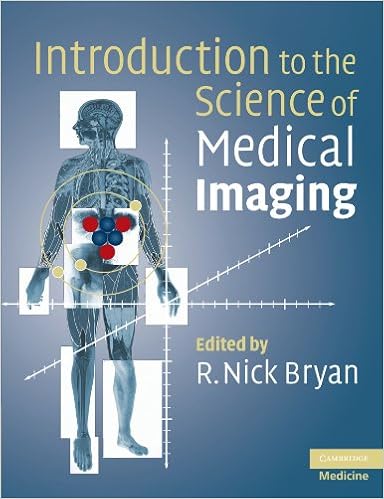
Introduction to the Science of Medical Imaging
Innovative advances in imaging expertise that supply excessive solution, 3D, non-invasive imaging of organic matters have made biomedical imaging a necessary software in scientific medication and biomedical study. Key technological advances comprise MRI, positron emission tomography (PET) and multidetector X-ray CT scanners.
- Cardiac imaging: a multimodality approach
- Textbook of Clinical Neurology, Goetz issue Neurology
- Ultrasound: a practical approach to clinical problems
- Evidence-Based Imaging in Pediatrics: Optimizing Imaging in Pediatric Patient Care
Additional info for 3D Angiographic Atlas of Neurovascular Anatomy and Pathology
Sample text
In Neuroimaging. W. Orrrison, ed. B. Saunders, pp. 206–212. 3. Borden NB, D. L. A. Flom. 1996. Postraumatic epistaxis from injury to the pterygovaginal artery. Am. J. Neuroradiol. 17:1148–1150. 4. E. Rosenbaum. 1974. The normal and anomalous aortic arch and brachiocephalic arteries. In Radiology of the Skull and Brain: Angiography Volume 2. H. G. , St. V. 1145–1163. 47 3D ANGIOGRAPHIC ATLAS OF NEUROVASCULAR ANATOMY AND PATHOLOGY 5. Berenstein A, and P. Lasjaunias. 1992. Surgical Neuroangiography: Functional Anatomy of the Craniofacial Arteries, Volume 1.
The ipsilateral parieto-occipital region has been removed. The medial surface of the contralateral parieto-occipital region is viewed. Also note the sylvian branches (45) of the ipsilateral middle cerebral artery overlying the insular cortex. The ipsilateral temporal lobe has been removed. Reprinted with permission of The Cleveland Clinic Foundation. 2 P2 segment of posterior cerebral artery 8 posterior temporal branch of posterior cerebral artery 9 parieto-occipital branch of posterior cerebral artery 10 calcarine branch of posterior cerebral artery 13m medial posterior choroidal artery 13L lateral posterior choroidal artery 14 vertebral-basilar junction 15 posterior pericallosal artery (splenial artery) 16 pontine perforator 17 anterior spinal artery RED KEY 10 supraclinoid (C2) segment internal carotid artery 12 posterior communicating artery 45 sylvian(insular) branches of middle cerebral artery 23 3D ANGIOGRAPHIC ATLAS OF NEUROVASCULAR ANATOMY AND PATHOLOGY Fig.
Lasjaunias. 1992. Surgical Neuroangiography: Functional Anatomy of the Craniofacial Arteries, Volume 1. New York: Springer-Verlag, 1992, pp. 239–244. 6. Berenstein and Lasjaunias, pp. 231–237. 7. L. Stillwell. 1969. Arteries and Veins of the Human Brain. Springfield, Ill: Charles C Thomas, pp. 71–73. 48 CERVICAL VASCULATURE (a) (b) Fig. 1 (a, b) Frontal (a) and lateral (b) left common carotid artery injection. Normal 2D appearance of the left common carotid artery bifurcation region. FIGURE KEY (a) 1 common carotid artery 2 internal carotid artery 2b carotid bulb 3 external carotid artery 4 ascending pharyngeal artery 5 occipital artery 6 posterior auricular artery 7 superior thyroid artery 9 lingual artery 10 facial artery 11 superficial temporal artery 12 internal maxillary artery (b) 49 3D ANGIOGRAPHIC ATLAS OF NEUROVASCULAR ANATOMY AND PATHOLOGY (a) (b) (a) (b) Fig.